KNEE CARTILAGE REPAIR PERTH

Knee Cartilage Repair Surgery – A patient’s guide:

 Australian Academy of Robotic
Orthopaedic Surgeons 

Knee Cartilage Repair Perth

Access the buttons below to be taken to the relevant information about knee cartilage repair surgery:
“We aim to provide you with personalised and unique medical care and ensure you fully understand your diagnosis and treatment options.”

Please feel free to ask any questions and bring a family member or friend along for support.

Australian Academy of Robotic Orthopaedic Surgeons
Robotic Orthopaedic Surgery Specialists.

What is Knee Cartilage

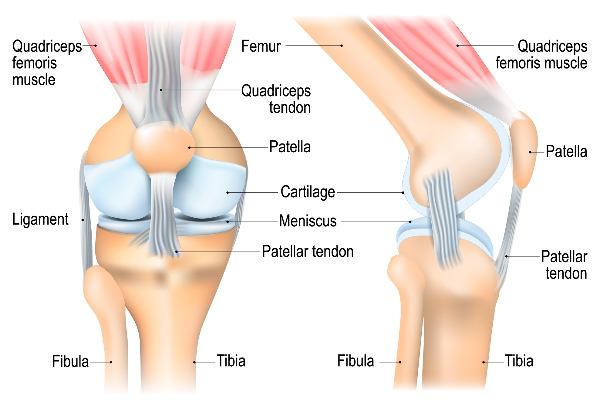
Your knee joints are lined by extremely smooth tissue called cartilage.

The cartilage of the knee joint lines:

  • The end of the thigh bone (or femur)

  • The top surface of the shin bone (or tibia) and;

  • The rear surface of the kneecap (or patella).

The cartilage is a protective cushion, allowing smooth movement of the joints as one bone end moves on the other.

Cartilage tissue covers the adjacent bone surfaces of the knee.
Knee Cartilage Repair Surgery – Aim

The aim of knee cartilage repair surgery is to:

  • Reduce pain

  • Improve joint function and;

  • Prevent further damage to the knee joint.

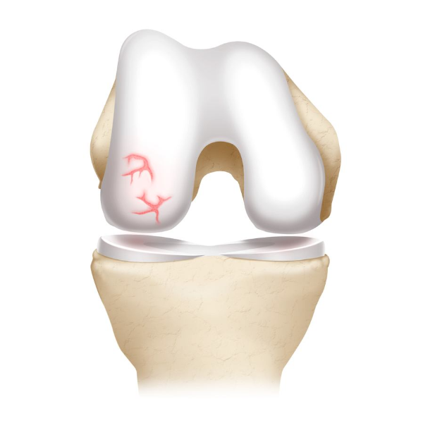
The size and location of the cartilage damage (or lesion) in the knee joint and the status of the other structures within the knee joint will help determine whether surgery is possible/suitable for your unique diagnosis.

Other structures within the knee may need to be assessed including, knee alignment, ligaments, cartilage damage or other factors.

These assessments will help identify patients who are more or less likely to benefit from the surgery and help determine the best surgical approach.
Knee Arthroscopy Image

Further knee joint assessment

Further investigations of the knee joint may reveal other structures in need of removal, trimming or repair including:

  • Removal of loose fragments of bone or cartilage within the knee

  • Trimming of damaged cartilage

  • Removal or repair of a torn meniscus

  • Removal of inflamed synovial tissue

  • Reconstruction of a torn anterior cruciate ligament

  • Treatment of patella (kneecap) problems

  • Treatment of knee sepsis (infection)

  • Identification of arthritis or osteoarthritis within the knee joint

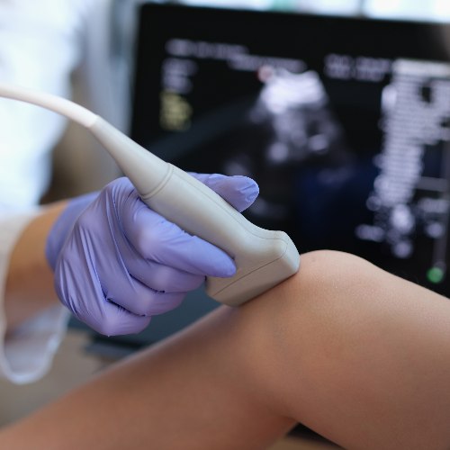
Further knee assessment tests may include x-rays, ultra-sound or MRI or CT scan.
Knee Arthroscopy Image

Types of Knee Cartilage Repair Surgery

There are five main types of knee cartilage repair surgery.

The benefits and risks of the most appropriate surgical approach to your unique situation will be discussed with you, by your surgeon to ensure you have all the information to make an informed decision about your diagnosis and treatment options.

Please access the buttons below to find out more about the main knee cartilage repair surgical approaches:

1. Chondroplasty

Chondroplasty is a surgical procedure performed on the knee to address damaged or degenerated cartilage. During the procedure, your surgeon uses arthroscopy, a minimally invasive technique that involves small incisions and a camera, to view the inside of the knee joint. Once the joint is accessed, the surgeon employs specialised tools to trim, reshape, or smooth the damaged cartilage surfaces.

This may involve removing loose or frayed cartilage fragments, repairing minor defects, or smoothing out rough areas. Removal of damaged tissue allows healthy cartilage to be able to grow in its place.

Because it’s a minimally invasive procedure, a chondroplasty requires fewer incisions than conventional surgery and patients may be able to recover more quickly than via an open surgical procedure.

2. Microfracture

Microfracture knee surgery is a minimally invasive procedure designed to treat small cartilage defects in the knee joint. Typically performed arthroscopically, this technique involves creating tiny fractures or “microfractures” in the underlying bone near the damaged cartilage. The purpose is to stimulate the growth of new cartilage by allowing bone marrow cells to reach the injured area.

During the procedure, the surgeon cleans the joint and identifies the cartilage lesion. Small holes or fractures are then made in the subchondral bone using a specialised tool. This process encourages blood flow and the release of bone marrow cells, which form a blood clot that eventually transforms into fibrocartilage.

While microfracture surgery can be effective in promoting the formation of new cartilage, the resulting tissue may not be as durable or resilient as the original.

Microfracture is a relatively common and successful treatment for small cartilage defects, it may not be suitable for larger lesions or in cases where the damage extends beyond the knee cartilage.

3. Autologous Minced Chondrocyte Implantation

Autologous Minced Chondrocyte Implantation (AMCI) is a cutting-edge knee surgery designed to treat cartilage defects and injuries. During the procedure, a small piece of healthy cartilage is extracted from the patient’s knee joint. Specialised cells called chondrocytes are isolated from this tissue and minced into tiny fragments. These minced chondrocytes are then implanted into the damaged area of the knee, promoting the regeneration of cartilage.

The surgery is performed arthroscopically, minimising invasiveness and reducing recovery time. AMCI offers a personalised approach, as the patient’s own cells are used, reducing the risk of rejection or complications. The minced chondrocytes stimulate the growth of new, healthy cartilage, addressing issues like osteoarthritis or focal cartilage defects.

AMCI is suitable for treating cartilage damage caused by direct injury (e.g. sports injuries, car accidents, falls) where the damaged area is surrounded by normal healthy cartilage.

Osteoarthritis and rheumatoid arthritic patients are not suitable for this treatment.

4. Matrix-induced Autologous Chondrocyte Implantation

Matrix-induced autologous chondrocyte implantation (MACI) technique involves harvesting a patient’s own healthy cartilage cells (called chondrocytes) from a non-weight-bearing area of the knee. These cells are then cultured and expanded in a laboratory.

During the surgery, the damaged cartilage in the knee is removed, creating a defect. A biocompatible scaffold is then implanted into the defect, serving as a framework for the cultured chondrocytes. The matrix helps the cells adhere and grow, facilitating the regeneration of new, healthy cartilage in the knee joint.

5. Osteochondral Autograft/Allograft Transplantation

Osteochondral autograft/allograft transplantation is a surgical procedure used to treat knee joint damage, particularly focal lesions or defects in the articular cartilage and underlying bone.

This technique involves the transfer of healthy cartilage and bone tissue from either the patient’s own body (autograft) or a donor (allograft) to the affected area of the knee.

During the surgery, the damaged cartilage and bone are first removed, creating a defect that is then filled with the graft tissue and bone.

This procedure is often considered for younger, active individuals with isolated cartilage lesions, as it aims to provide a durable and long-term solution for joint preservation.

Recovery from surgical procedures

Post-surgery rehabilitation is crucial for a successful outcome, involving a gradual return to weight-bearing activities and physical therapy to optimise healing and knee joint function.

The most appropriate approach to cartilage repair will be discussed with you by your surgeon. Individual outcomes from surgery vary. Please discuss with your surgeon to determine the most suitable treatment plan based on your specific condition.

After Knee Cartilage Repair Surgery

Recovery times may vary from patient to patient for a number of reasons including age, current heath, surgical complexity, muscle and bone strength and commitment to rehabilitation amongst many other factors.

Surgery – The surgery itself usually takes 1 – 2 hours.

Post-surgery – For some patients it’s day surgery, so you will be observed for a couple of hours to ensure that you have recovered from the anaesthetic and discharged from hospital.

Some patients will stay overnight in hospital.

After surgery you will probably feel a bit sleepy and might need some pain killers. Have a friend or family member available to help you for a few days after you leave hospital.

You will be given instructions on:

  • Wound care management and dressings

  • Warning signs to look out for

  • Pain management

  • Rehabilitation plan

  • Follow up appointments

If you have any questions, please contact the team at the Australian Academy of Robotic Orthopaedic Surgeons.
Robotic Orthopaedic Surgery Specialists.

After Surgery & Rehabilitation – Frequently Asked Questions:

Movement after surgery – Depending on the type of surgery undertaken your surgeon may initially recommend moving around with crutches for the first few weeks after surgery for comfort, safety and confidence and to give the knee time to heal and repair.

Physiotherapy recovery program – You will see a physiotherapist and be prescribed a rehabilitation program after your surgery. Prior to discharge you will be given exercise instructions and practice on how to best perform daily activities including climbing stairs, getting into and out of cars etc.

Will I need time off work – Returning to work depends on your occupation, duties and the procedure performed. Most patients can return to office work within one week. Labour-intensive work may require up to 6 weeks before returning to complete duties particularly if they involve prolonged standing, heavy lifting, bending or repetitive stair climbing.

Driving – Driving a car is not recommended for 48 hours after an anaesthetic. After the initial 48 hours, your ability to drive will depend on the knee you had your operation on (left or right), the vehicle you drive (manual or automatic), and the type of procedure you had.

Please confirm with your surgeon to discuss your situation in detail.

Air travel – Air travel may be undertaken soon after you are cleared by your surgeon.

Return to sporting activities – As the knee joint cartilage has been damaged, activities such as long-distance running are not recommended as this can result in rapid deterioration of the knee joint cartilage.

It is best to delay leisure activities or sports for six weeks to allow the cartilage to heal and repair. However, depending on what type of procedure is needed, more or less time off sports will be required. Your surgeon can provide more detail.

What are the Risks of Knee Cartilage Repair Surgery

Potential risks knee of knee cartilage repair surgery – Knee cartilage repair surgery is very safe, and serious complications are uncommon. The surgeries have a high success rate in reducing pain and improving knee function and mobility.

It may take several months to a year for the full benefits of the procedure to be realised. Some patients may need additional surgeries or treatments to manage symptoms in more complex cases.

As with any major surgery, there are potential risks involved. The decision to proceed with the surgery is made because the advantages of surgery outweigh the potential disadvantages.

It is important that you are informed of these risks before the surgery takes place. Complications can be medical (general) or local complications specific to the knee. Medical complications include those of the anaesthetic and your general well being. Almost any medical condition can occur so this list is not complete.

Complications that may include:

  • Allergic reactions to medications

  • Blood loss requiring transfusion

  • Heart attacks, strokes, kidney failure, pneumonia and bladder infections

  • Nerve damage

  • Ongoing health concerns, prolonged hospitalisation or rarely death.

  • Risk of infection

  • Blood clots

  • Wound irritation

  • Pain

If you have any questions, please contact the team at the Australian Academy of Robotic Orthopaedic Surgeons.
Robotic Orthopaedic Surgery Specialists.